Most Read
Abstract
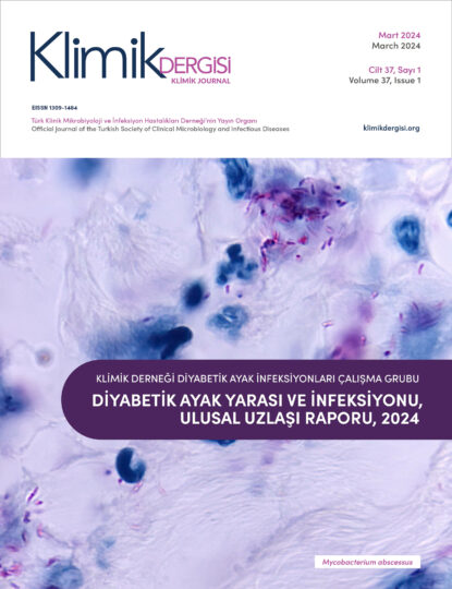
Study Group for Diabetic Foot Infections of the Turkish Society of Clinical Microbiology and Infectious Diseases has called for collaboration of the relevant specialist societies and the Ministry of Health to issue a national consensus report on the diagnosis, treatment and prevention of diabetic foot (DF) wounds and diabetic foot infections (DFIs) in Turkey. In the periodical meetings of the assigned representatives from all the parties, various questions as to pathogenesis, microbiology, assessment and grading, treatment, prevention and control of diabetic foot were identified. Upon reviewing related literature and international guidelines, these questions were provided with consensus answers. Several of the answers provided in the report are listed below: [1] Although there are many reasons for the development of DF wounds, the main reason is the combined effect of diabetes-related vascular disease and neuropathy. [2] Aerobic Gram-positive cocci are mostly responsible for superficial DFIs in patients with cellulitis and no history of antibiotic use. [3] Pseudomonas aeruginosa is one of the commonly encountered agents when between the toes of the patient are moist. [4] When the other potential reasons are eliminated, DFIs should be considered in presence of at least two of the classical signs of inflammation including redness, warmth, swelling, tenderness, and pain, or purulent discharge in the foot lesion. [5] Infections are classified into mild, moderate, or severe groups according to some criteria such as the depth and width of the wounds, and the presence of systemic findings of infection. [6] PEDIS system should be preferred as a classification system for its high predictive value in diabetes-related foot complications. [7] Culture samples from the DF wound should only be obtained when infection is clinically considered and, where possible, before starting antibiotic treatment. [8] Inflammatory biomarkers such as leukocyte count, C-reactive protein, erythrocyte sedimentation rate, and procalcitonin may be useful in distinguishing between colonization with infection. [9] Magnetic resonance imaging is a sensitive and specific method in patients unresponsive to treatment when osteomyelitis and deep soft tissue abscesses are considered. [10] The gold standard in the diagnosis of osteomyelitis is histopathological examination. [11] To provide wound healing and to save the limb, removal of dead and infected tissue with urgent and aggressive debridement, appropriate antibiotic therapy, metabolic control, and off-loading of pressure, the diagnosis and proper treatment of peripheral arterial disease, and restoration of the foot function are necessary. [12] A lot of different factors playing a role in etiopathogenesis complicate the approach to be developed in this type of lesions, and therefore it requires a team concept. [13] In the empirical treatment, the objective should be treating only the potential agents. Adequate tissue levels, low side effects and patient compliance must be observed; effective drugs should be used in specified doses and duration. [14] Debridement is an essential and integral part of wound treatment and is an important tool allowing the formation of healthy granulation tissue. [15] When the infected tissue cannot be completely cleared with the debridement and in cases when the patient could not cope with the remaining infection load, performing a limb amputation on a safe level of infection would be lifesaving. [16] If an arterial insufficiency is considered in a patient with a DF wound, early diagnosis and interventional treatment is necessary. [17] Hyperbaric oxygen therapy is used as an adjunctive treatment in combination with other treatments in DFI patients. [18] Topical negative pressure therapy is a useful adjunctive measure in selected patients. [19] Growth factors can be used in selected patients other than wounds that can be treated with cheaper and safer methods. [20] Maggot therapy may be considered as a debridement method in DF wound cases. [21] Patients with more than ten years of diabetes history have an increased risk of wound development or amputation. [22] DF problem is the only complication of diabetes that can be prevented through education.
