Most Read
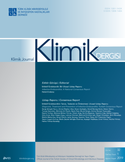
Although infective endocarditis (IE) is rare, it is still important as an infectious disease because of the resulting morbidity and substantial mortality rates. Epidemiological studies in developed countries have shown that the incidence of IE has been approximately 6/100 000 in recent years and it is on the fourth rank among the most life-threatening infectious diseases after sepsis, pneumonia and intraabdominal infections. Although IE is not a reportable disease in Turkey, and an incidence study was not performed, its incidence may be expected to be higher due to both more frequent presence of predisposing cardiac conditions and higher rates of nosocomial bacteremia which may lead to IE in risk groups. Additionally, while IE generally affects elderly people in developed countries it still affects young people in Turkey. In order to reduce the mortality and morbidity, it is critical to diagnose the IE, to determine the causative agent and to start treatment rapidly. However, most of the patients cannot be diagnosed in their first visits, about half of them can be diagnosed after 3 months, and the disease often goes unnoticed. In patients diagnosed as IE, the rate of identification of causative organisms is more than 90% in developed countries, while it is around 60% in Turkey. Furthermore, some important microbiological diagnostic tests are not performed in most of the centers. Some antimicrobials that are recommended as the first option for treatment of IE, particularly antistaphylococcal penicillins, are unavailable in Turkey. These problems necessitate to review the epidemiological, laboratory and clinical characteristics of IE in the country, as well as the current information about its diagnosis, treatment and prevention together with local data. Patients with IE can be followed by physicians in many specialties. Diagnosis and treatment processes of IE should be standardized at every stage so that management of IE, a setting in which many physicians are involved, can always be in line with current recommendations. From this point of view, Study Group for Infective Endocarditis and Other Cardiovascular Infections of the Turkish Society of Clinical Microbiology and Infectious Diseases has called for collaboration of the relevant specialist organizations to establish a consensus report on the diagnosis, treatment and prevention of IE in the light of current information and local data in Turkey. In the periodical meetings of the assigned representatives from all the parties, various questions were identified. Upon reviewing related literature and international guidelines, these questions were provided with consensus answers. Several of the answers provided in the report are listed below: [1] IE is more frequent in patients with a previous episode of IE, a valvular heart disease, a congenital heart disease, any intracardiac prosthetic material, an intravenous drug addiction, chronic hemodialysis treatment, solid organ and hematopoietic stem cell transplantation as compared with normal population. [2] The most frequent causative organisms are Staphylococcus aureus, streptococci, coagulase-negative staphylococci, and enterococci, respectively, both in Turkey and globally. Brucella spp. is the fifth common causative agent of IE in Turkey. [3] The echocardiograpghy is the imaging modality of choice to define cardiac lesions in patients with suspected IE. Both transthoracic and transesophageal echocardiography are generally necessary in almost all patients. Both are inconclusive approximately in 15% of total IE cases whereas the percentage is up to 30% in patients with intracardiac prosthetic devices. In these instances, multi-slice (MS) computed tomography (CT) should be the imaging modality in patients with native valve IE, whereas MS-CT or radiolabelled leukocyte scintigraphy with single-photon emission tomography/CT should be choosen for patients who have prosthetic valve IE within the first 3 months of surgery, and MS-CT or positron-emission tomography/CT should be chosen for patients with prosthetic valve IE after 3 months of surgery. [4] Blood cultures should be taken without any delay to catch-up the febrile period as 3 sets with 30-minute intervals (3 aerobic and 3 anaerobic bottles, totally 6 bottles) in patients with suspected IE. Each set, comprised of 1 aerobic and 1 anaerobic bottle, should be inoculated with 18-20 ml of blood (9 -10 ml blood per bottle). Totally 60 ml of blood should be taken from one patient with suspected IE. Two sets of control blood cultures should be repeated in every 48 hours after initiation of therapy in order to show blood sterility. If causative organism do not grow in the usual blood culture bottles, additional three mycobacterial blood culture bottles should be inoculated in patients with suspected prosthetic valve IE and who had a cardiac surgery in the last decade. [5] The excised valvular tissue from patients with suspected IE should be evaluated both microbiologically and histopathologically. [6] First of all, Wright agglutination test (if negative, by adding Coombs’ serum) and indirect fluorescent antibody (IFA) test to investigate Coxiella burnetii phase I IgG antibodies should be done in culture-negative patients. If these two tests are negative, IgG antibodies for Bartonella spp., Legionella spp., Chlamydia spp., and Mycoplasma spp. should be tested respectively and preferably by IFA test. [7] Multiplex polymerase chain reaction (PCR) tests should be used to identify the pathogen in whole blood in a culture-negative patient who has received previous antibiotic therapy. If the blood cultures are negative in a patient who has not received previous antibiotic therapy, PCR tests for 16S rRNA gene analysis and Tropheryma whipplei should be performed on the resected valve obtained during surgery. [8] Histopathological examination of resected valvular tissue in patients with suspected IE give valuable information about the activation and degree of the inflammation. Moreover, histopathological examination with appropriate routine and immunohistochemical staining, aid to identify especially intracellular pathogens like C. burnetii, Bartonella spp. and T. whipplei in blood culture-negative patients. [9] Bactericidal agents given parenterally for long duration is the general principle of antimicrobial treatment of IE. The pathogenic organism, presence of prosthetic material and duration of symptoms specifies the duration of treatment. The therapy duration is generally 4-6 weeks for native valve IE and >6 weeks for prosthetic valve IE. [10] As the efficacy and feasibility of oral antimicrobial choices of left-sided IE are not well defined in Turkey and it is related with substantial mortality, parenteral route should be preferred for the complete duration of antimicrobial treatment of left-sided IE in Turkey. In case of unavailability of intravenous access or outpatient parenteral antibiotic therapy, oral agents may be feasible to complete the therapy duration in stable patients with uncomplicated native valve IE due to drug-susceptible viridans streptococci, provided that initial two weeks should be completed parenterally, and the patient should give an informed consent after notifying all possible risks, and regular post-discharge follow-up should be possible. The decision for oral maintenance therapy has to be given by the IE team. [11] The appropriate antimicrobials should be initiated without any delay as it reduces not only the risk of an embolic event in patients with either acute or subacute IE, but also decreases the mortality associated with sepsis in acute IE. Therefore, the empirical antimicrobials should be promptly initiated after blood cultures are taken. [12] Ampicillin-sulbactam ± gentamicin can be initiated empirically in the treatment of community-acquired, both acute and subacute types of native and late prosthetic valve IE in adults whereas either vancomycin + ampicillin-sulbactam or ceftriaxone ± gentamicin can be the choice for acute types. Vancomycin + cefepime ± gentamicin combination can be initiated empirically in the treatment of nosocomial native, early and late prosthetic valve IE in adults. Gentamicin should be avoided initially in patients with impaired renal function. Rifampin can be added to initial empirical treatment of early prosthetic valve IE. Daptomycin alone is not a drug of choice for initial empirical treatment of IE because of its suboptimal efficacy for streptococci and enterococci in which resistance can easily develop during therapy.
Klimik Dergisi 2019; 32(Suppl. 1): 2-116.
Cite this article as: Şimşek-Yavuz S, Akar AR, Aydoğdu S, et al. [Diagnosis, treatment and prevention of infective endocarditis: Turkish consensus report]. Klimik Derg. 2019; 32(Suppl. 1): 2-116. Turkish.
